Remove hearing aids before panoramic dental x-ray?

Thanks for your efforts to find the answer. I suspect that the actuaries and lawyers have gotten together and decided to include a warning because:

  1. X-rays are becoming more ubiquitous for security reasons, and

  2. Chip foundries are using smaller and smaller architecture (Oticon’s new Polaris chip is 28nm - small for HAs, I’ve read).

Still, I am convinced that accidental leakage of strong EM flux fields is a real threat to some electronic devices - hearing aids among them.

I may be frightened of this Phantom EM/RF Monster for no good reason, but every time I cook something in my microwave oven, I wonder.

À bientôt
Spud

I asked this question to the Dutch Institute of Clinical Physics, here is their answer:

Dear Mr. XXXXX

I submitted your question to our Radiation Hygiene Committee and received the following answer:

From a physical point of view we cannot imagine that a hearing aid can break, but we cannot give individual advice and the patient should consult his / her audiologist / hearing care professional for this. We can say that with regard to the image quality a hearing aid may affect when an image is taken in this region. We recommend that you discuss this with the appropriate physician or employee in the imaging department conducting the examination.

Sounds like sort of a non-answer, doesn’t it?

I wish there was more of a definite answer. I have a small salivary stone in the left maxillary salivary duct. It moves around, so sometimes the dental surgeon takes a panoramic view x-ray.

I don’t want my wonderful new Oticon devices to end up as More Smoke!

If you are worried, take them out. I had a pan at my dentist yesterday and they said to remove them, but I doubt it was really necessary.

1 Like

Apparently, the hearing aids often cause shadows and other artifacts when worn during a panoramic dental X-ray. Nothing to do with frying the devices.

At least that’s my understanding…

==============================
[EDIT 22April/'21: I had a dentist appointment yesterday and I asked him about hearing aids and X-ray radiation. He confirmed that wearing hearing aids during panoramic X-rays can cause imaging problems, but he’s never heard of the hearing devices being damaged.

DrMike is well-read (i.e., he keeps current with journals and scholarly articles). I trust what he’s telling me …]

My wonderful Veterans’ Affairs Canada :canada: 
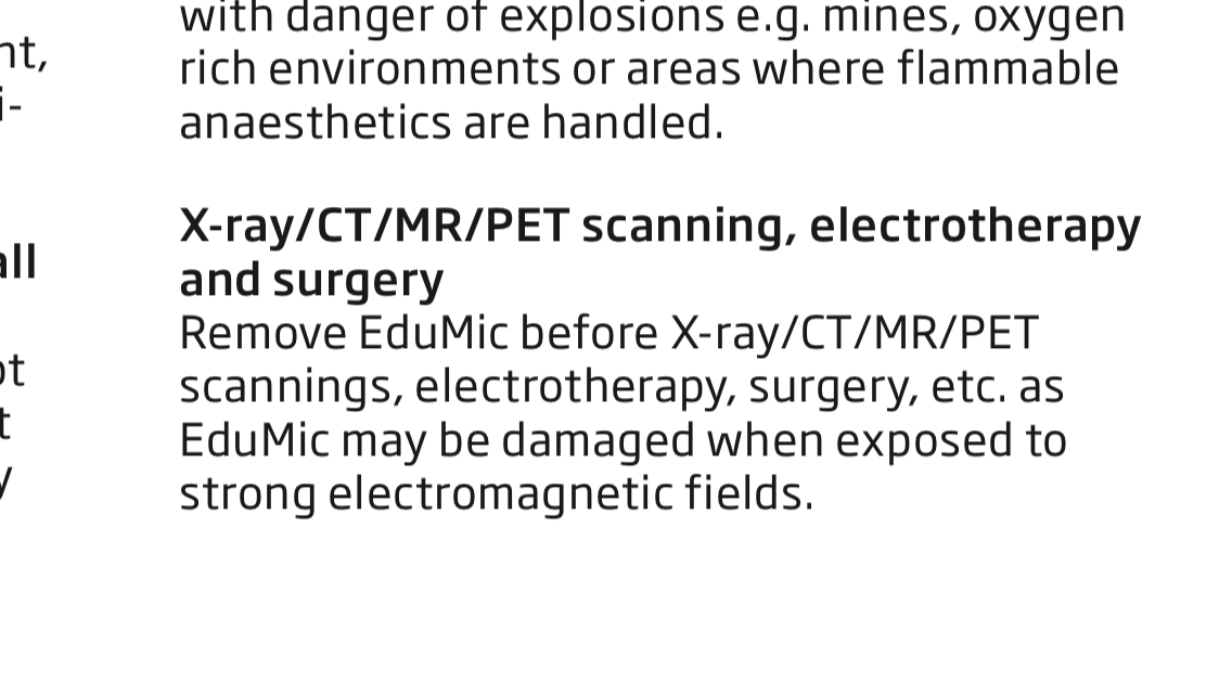
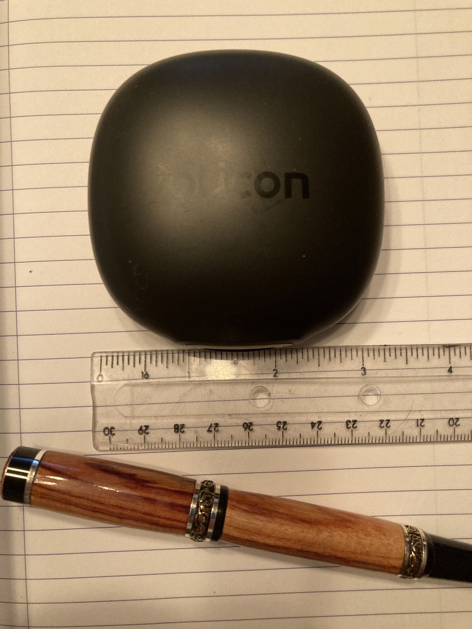
has just approved me for an Oticon EduMic.

I was reading the online manual, which says this:

I don’t see why/how the HAs themselves would be any less susceptible to damage than the EduMic.

FYI

1 Like

Long ago I involved in this topic and some time ago I got some new information from the Dutch productmanager from Signia (WS Audiology: Signia and Sivantos). I would like to add it here to inform you about my research. This is what I got from him (thanks to Google translate for the English translation):
It appears that X-ray examination can affect the fine structures of NVM memory circuits. This is the permanent memory in hearing aids. Tests are done with the hearing aids and X-rays with a power of 100kV/100mGy. More than 100kV can potentially damage the chips, hence the warning in the manual for hearing aids. Should the hearing aids be damaged, this will have no further consequences for the safety or health of the hearing aid user. If damage occurs, the hearing aid may no longer meet the requirements for processing and processing the sound, causing the hearing aid user to notice that the hearing aid is not functioning.
(fyi Panoramic dental x- ray is far beneath 100kV)

1 Like

Speaking as an EE, with a Masters in microelectronics technology, and a career designing chips, I can assure you that exposure of memory chips (Eprom, EEprom, and Flash) to X rays can cause the miniscule amounts of stored charge inside them to dissipate.

Potentially blanking out or just corrupting the data inside them. Firmware, custom programs etc. Even if not immediately damaged, weakened and liable to corrupt much sooner than otherwise. Why take the risk ? Why fixated on keeping your aids in during any such procedure ?

Zap such a memory chip enough, to corrupt the bootloader, and the HA manufacturer will not be able to recover it. Except maybe by de-soldering it. Which is hardly a practical possibility.

CT scans, or other significant doses from medical X rays, have the potential to zap the memory chips. Even if they don’t at first, they can weaken the charge inside, sometimes only a few electrons.

I have done that before, to blank out otherwise on-time programmable processors or memory chips. which were plastic encapsulated, not ceramic with a window in them to zap them with UV light.

MRI’s are different, no ionizing radiation but extreme magnetic and RF fields. Also very capable of destroying electronic circuitry by quite different mechanisms…

No competent radiologist would allow you anywhere near an MRI machine with hearing aids in your ears. I have had three now, and they were even concerned as to whether I might have some tattoos or even piercings (no). Apparently even some tattoo ink can heat up in that environment and cause skin burns. I do have some bits of titanium inside my ears which they knew about, and knew were not going to be a problem.

Knowing that I have also worked in an industrial environment, potentially exposed to shards of metal getting into my eyes, happened once but extracted, they were also concerned about that, as well as the fact that I eat game, shot with steel shot, some of which might still be inside my bowels. But head MRIs only, so they decided not to CT me first to look out for that.

Frankly, the HA manufacturers do know, when they caution against allowing their aids to be exposed to e.g. medical X-rays. Of which CT scans are quite a powerful variety. Why on earth would you do so, for no good reason ?

Luggage screening at airports etc. is at a far lower level, nothing too much to worry about with that.

2 Likes

Thank you very much Surdi for your reply. By searching for someone in this field of knowledge… I appreciate very much you input. The topic though is “Remove hearing aids before panoramic dental x-ray.”

So as far as I understand from the productmanager from Signia he says Tests are done with the hearing aids and X-rays with a power of 100kV/100mGy. More than 100kV can potentially damage the chips”.
You can assure this also, thats fine. But unfortunately you do not give any specification. So what is your opinion about the specified kV and mGy as that is the important detail missing.
I have been an academical radiologist myself and started on MRI and CT’s at the very beginning. So I have some basic knowledge, as a radiation expert, about X-rays, ultrasound and magnetic resonance, but not about memory modules in HA’s.

Of course, as you also state, magnetic metal in an MRI is not allowed! Thats why every MRI institute will ask questions like did you work in metalindustrie or are you aware of metal- implants which could be even tatoo- ink, and makeup for camouflage of skintumors or even makeup for whatsoever reason and so on!. And these can even lead to faildiagnoses even in x- ray examinations.

The kV used by panoramic dental x-rays is not in the range of 100kV and over. So the question for me stays what about the low kV in panoramic dental x-rays specific. That will be about 70kV… Maybe you can give some information about that I hope.

About your question “Why take the risk ? Why fixated on keeping your aids in during any such procedure ?” Sometimes patients arrive in the x- Ray dept. in f.i. emergency cases or you want to give them instructions during an examination they have to hear you. So my perspective is a bit different then that of the patient I can have a good conversation with. Nowadays there are HA’s wich are in the canal and not easily visible anymore in a fast inspection. And most of the time we have to ask a patient more than just if he wears HA’s. Thats why I am personaly so persistent in knowing exactly what is important to not damage HA’s.

1 Like

I have been in this situation recently (unexpected/unexplained arrhythmia and high BP). My regular HAs were out of charge when the arrhythmia hit, so I was obliged to wear my spares, which are more or less useless to me.

The interruptions in the dialogue with my medical caregivers caused by my inability to understand questions and instructions were frustrating, annoying, and potentially dangerous!

Fortunately, our hospital clearly labels forms, files - and even my person - with big blue HoH stickers, which somewhat prepare staff for the difficulty we will have communicating.

My HAs came with an easily-pocketable case, so that somewhat facilitates the logistical problems of moving around the hospital with HAs in tow (you can even duct tape a loop of paracord to the case to fashion a temporary lanyard). Too bad I still was unable to understand with the devices I was toting around!

IMO, HAs are essential during medical exams/procedures.

2 Likes

emile.heilbron, I am not familiar with the characteristics of medical x-rays and CT scanners, being more concerned from an electronics design and manufacturing perspective.

There has been lots of work done looking into the effects of x-ray PCB inspection methods, where the modern very small geometry Flash devices nowadays ubiquitous, have been found to be very susceptible to the effects of x-rays.

After a quick search, I have come up with a few links that may be of interest:

X-ray irradiation was performed using an ARACOR Model
4100 10-keV X-ray irradiator at a dose rate of 5 krad(SiO2)/
min.

The total ionizing dose response of a triple-level-cell (TLC) NAND flash is shown to be low enough that data corruption can occur as a result of an x-ray inspection. Only a few seconds of x-ray exposure corresponding to a total dose of merely 50 rad(Si) in a real-time x-ray source are required to induce errors.

https://meridian.allenpress.com/ism/article-abstract/2016/1/000660/187897/Effects-of-x-ray-exposure-on-NOR-and-NAND-flash

In this paper, we present a detailed study on the effects of x-ray exposure on data corruption in commercially available NOR and NAND flash memory devices during x-ray inspection with a high-resolution Phoenix Nanomex system from GE. We investigated role of the x-ray tube voltage, tube current, device orientation, x-ray filters and photon energy.

The dosimetry in these studies is usually reported in rads of Silicon equivalent. I do not know how that compares with the methods adopted for medical X-rays, which I suppose assume a different ,material for bodily absorption. But, for example, the ieee paper reports that doses as low as 50 rad(Si).(i.e 50 cGy(Si) can cause corruption.

Such damage is cumulative. I.e. a chip may survive one exposure of a certain dose, but repeat exposures may steadily weaken it until the data does become corrupted.

Apparently the plastic packaging of discrete components has some shielding effect on x-rays of below 10 KV, but that protection was found to be removed when some chips were de-capped before the tests. And will of course depend on the thickness of the plastic layer. I don’t know much about the construction of the actual chip assemblies inside modern hearing aids, nor whether the outer plastic casings might themselves have some shielding effect.

I came across a table here: Radiation risk from medical imaging - Harvard Health from which it seems that the dose from one panoramic dental x-ray is about ten times higher than that from say an arm or leg x-ray (0.01 mSv vs. 0.001 mSv)

Whereas a head CT scan could be 2 mSv. Or more.

NB. these are averages, if you look at that table they also give the ranges reported in studies, which vary considerably.

Having had three head CT scans in the past year, as well as a full cerebral angiogram, and a barium swallow test, I am becoming slightly nervous about having too many more high dose procedures.

My guess is that if anything is going to zap hearing aids, it would be having a head CT scan whilst wearing them. I have no knowledge of panoramic dental x-rays, mine are done as individual ones.

I am sorry if that is not terribly helpful, but there is research out there to find.

2 Likes

emile.helbron

Regarding variation of sensitivity to damage vs. x-ray voltage, you may find the following publication of interest. Table 2.0 refers.

But these tests were done on plastic packaged devices (thin quad flat pack), where the plastic has been observed in other studies to provide some shielding effect at lower voltages. As I said, I am not familiar with how modern hearing aid chip assemblies are packaged, but some of them might even be using bare chips, wirebonded to a ceramic substrate that also includes the electrical circuitry.

One study, using monochromatic synchrotron radiation, found that the peak sensitivity of the devices they were testing was at the 125 kV level. Which I think is where CT scanners often operate.

I could only find data about medical x-rays considering overall body exposure, in mSieverts. What matters to electronic devices is the actual flux through the part, times the time exposed, measured in rads/cGy, and based on the material’s capture, i.e. silicon for most (but not all) chips, and something else for human tissue (mostly water ?). I have not looked up the differences yet.

1 Like

That, might I suggest, is a little bit disingenuous. It might represent what they sometimes use for product inspection, but not necessarily what might happen during some medical procedures.

Over to you.

Meanwhile, from my experiences with three head CT scans, three head MRIs, a cerebral angiogram, and more, I have always been provided with headphones with mics. to communicate throughput the procedures, presumably specialised devices, at least for the MRIs, I had assumed that this was normal. The most recent ones (CT and MRI), the machines had loudspeakers and mics. built in, to communicate. In the MRI scanner I was given passive hearing protectors to quieten down the noises, which I found rather interesting, and not at all scary. Apparently some can freak out when having an MRI, so I suppose it is routine to offer them.

Otherwise, for routine consultations I keep my “ears” in.

1 Like

Thank you SpudGunner for what I consider to be a valuable contribution to this discussion in which you point out the sometimes conflicting interests that need to be weighed up.

For the more factual explanation I am happy with the information Surdi provides in his posts.

1 Like

@emile.heilbron: Thank you for your appreciation of my desire to contribute to your important topic. I am simply grateful that you and other learned members are actually giving this important subject some attention.

Thank you for keeping this topic alive, Doctor.

1 Like

Thank you surdi for your last three contributions and the substantiation through the supplied very relevant and interesting publications. I needed some time to read them carefully. An extensive discussion is possible on this to go into detail on these pieces. Probably more suitable for a dialogue among us nurds with Zoom or Skype or scientific publications than under this topic, which was started by mazda4me and asks for the answer to the more practical question “Remove hearing aids before panoramic dental x-ray?”. But personally I am very happy with your information.

The scientific value of this cannot be underestimated. The implications that the interface has among technical developers and users cannot be endorsed enough. The practical value for the HA user and audiologist and the user should be paramount here.

The X-ray beam used in a dental panoramic x-ray is a very special one compared to a CT and/or conventional examination. A special kind of ‘tomographic imaging’ is used. A cut slice with a depth of field/focus of a few centimeters centered along the line of the teeth. Various techniques are used to achieve the correct flat kV by means of tube voltage and focusing, including filters. And the exposure time is also very limited locally along the row of teeth. Of course, earrings, pearcings and HA are generally removed.

In practice this would mean that the radiation damage to the HAs is present. However, this is also unavoidable in nature: think of the use of aircraft at height and radiation from buildings in which we live.
As is also apparent from the articles you quoted, the Fail Bit Ratio of the memory used by the HA is undesirable, but does not necessarily have to lead to loss of function. However, it cannot be ruled out and has not to be ignored.

Even if you take the HA off for an OPT/dental panoramic x-ray, where would you like to put them safely in an x-ray dept at that moment? The same applies to your mobile phone, your creditcard and all other goods containing these chips.

By the way Surdi to go into one of your questions Computed Tomography exists in many forms. Multiple beams, but also multi energy/kV and automatic current adjustment. The more money, the more setting options in general.

In conclusion: If you are unlucky, your HAs will eventually break with or without dental panoramic x-ray. The chance this will happen is not very big, but do not take unnecesary risks. You will most of the time be asked to take them off because they can negatively influence the diagnostic value due to the disturbed imaging. In addition, this examination/potential risk is not done without a solid indication.

@emile.heilbron: I’m chuckling at the post I’m about to upload to the Forum! The image is one that shows technology at the other end of the spectrum of sophisticated diagnostic imaging devices that you scientists are discussing. It shows the standard issue Oticon carry case for More1 hearing aids…

This little case has a smooth, flat profile that makes it easy to pocket. The gowns you make us wear for diagnostic imaging procedures are without pockets, however, so I have successfully fashioned makeshift lanyards for my case by simply GorillaTaping a suitable length of paracord (with knots tied in each end) to the underside of the case. While this getup can’t be kept on one’s person during the actual operation of the X-ray source, it is unobtrusive enough to be able to be smuggled into examination rooms so that it’s close at hand when the moment comes when one’s hearing devices must be applied or removed.

My objective in designing the SpudCap(sule)®️ was not to subvert hospital protocols, but rather, it was to ensure the proximity and accessibility of my hearing aids, when they were needed in order to dialogue with my caregivers.

[During the ER visit to which I referred in my previous post, I presented doctors with a rather different set of facts. I had recently had a cortisone injection to my sacroiliac joint. The injection must have come too close to a nerve, and resulted in such intense pain that it precipitated an arrhythmia (right bundle branch block), and abnormally high blood pressure. The DD was uncommon enough that a rather long medical interrogation was necessary, during which I was obliged to wear my hearing aids, which had never been properly fitted to give me good comprehension. The frustration on the part of my caregivers was so pronounced that a nurse actually asked me if my aids were working. My inability to understand and answer questions on the first pass made me feel old, dull-witted, and disabled.][No imaging was involved in this episode, but I was prepared!]

My point is that medical professionals should not require me to check my hearing prosthetics at the door. They are part of me, without which I cannot function, and it’s no more appropriate to treat these as “checked baggage” as it would be to ask a patient to leave their prosthetic leg at the front desk.

The knowledge demanded of mankind today is so overwhelming that it is hard to comprehend :wink:

2 Likes

… my invention reflects my Neanderthal roots!

1 Like

I had a bilateral cataract removal operation recently. There is far too much liquid around for wearing hearing aids to be possible in that situation, so I took them out and put them into my pocket as I sat on the operating table.

I had already agreed with the surgeon that I would do this and said that I would need him to speak up if he wanted to ask or tell me something, in particular about whether to proceed with the second eye once the first was done. He said “That’s ok. I will shout. I need the exercise!”

2 Likes